 |
|||||||||||
|
|||||||||||
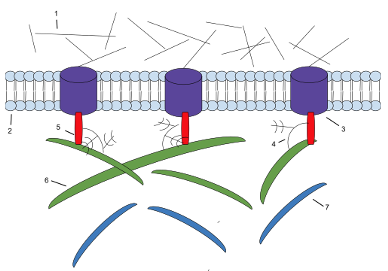
However, your cells mostly consist of water and so do all the tissues and organs in your body. So, how do they maintain their shape and what gives them structural and mechanical support? As a prior article (ECS-11) noted it is the cell’s cytoskeleton (microtubules, microfilaments and intermediate filaments) that give it shape and structural and mechanical support. And it is the connective tissue, consisting of cells (mostly fibroblasts) that secrete a gel-like ground substance and protein fibers, that crisscross through it, which provide your body’s tissues and organs with structural and mechanical support. The ground substance and protein fibers are called the extracellular matrix (ECM) which is the non-cellular component of connective tissue. In fact, different types of connective tissue provide different types of support. In the human body this runs from solid bones, to softer and more elastic cartilage, to high tensile strength ligaments and tendons, to the delicate web-like laced spider-like networks (bubble wrap) that supports most of its organs and passageways. It all depends upon the different types of cells which secrete different types of ground substance and the density and material qualities of the different protein fibers running through them. But the ECM does much more than just provide the body’s tissues and organs with structural and mechanical support. It also affects cell signaling, migration, growth, proliferation, differentiation and survival, all of which regulates tissue morphology, development, homeostasis and function. The last four articles in this series outlined how the ECM manages these functions through its main components; collagen and elastin fibers (ECS-12), the gel-like ground substance made of water, glycoaminoglycans (GAGs) and proteoglycans (PGs -ECS-13), glycoproteins (GPs), and cell surface receptors like integrins (ECS-14), and growth factors (GFs), cytokines, chemokines, matrikines, enzymes and their inhibitors, (ECS-15) (see Figure 1).
This article will look at how all the parts of the ECM (and more) work together in a coordinated fashion to accomplish wound healing. Keep in mind, that we are supposed to believe that an unguided process, like natural selection acting on random variation, was responsible for the presence of each of these components and what they in combination do for the body. A Symphony in Four Parts Wound healing is the coordinated, sequential, and overlapping set of biological processes that restore tissue integrity after injury. It can be divided into four stages:
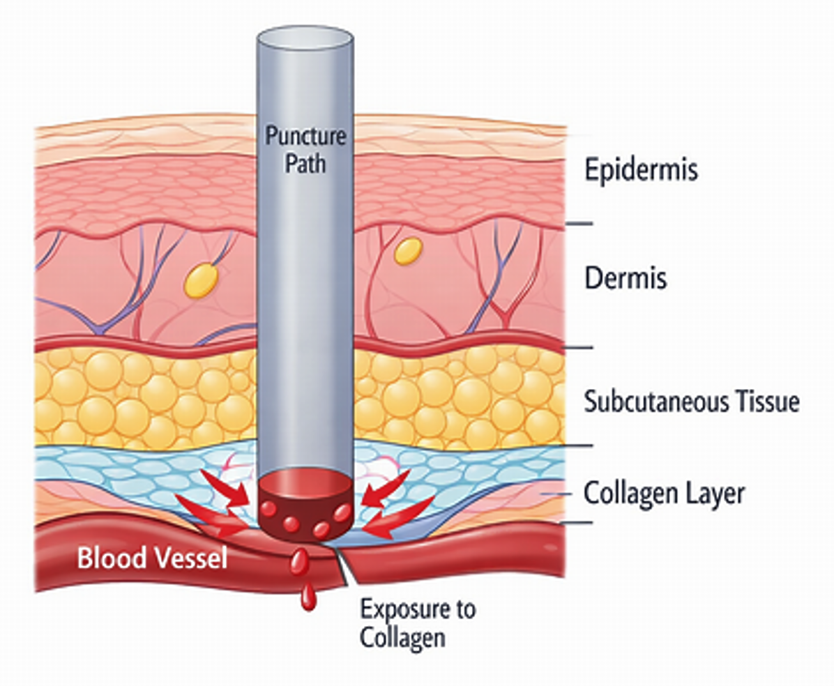
The order is functionally necessary; if any stage fails or activates at the wrong time, the tissue environment becomes chaotic and wound healing becomes impossible. Hemostasis is immediate, beginning within seconds and lasting for many minutes. The first response is rapid vasoconstriction, which reduces local blood flow and limits further blood loss. Platelets adhere to exposed subendothelial collagen and aggregate to form a temporary platelet plug, while fibrin polymerization stabilizes this plug into a firm clot. Together, these actions stop the bleeding and create the provisional matrix and signaling environment that initiate the next three phases in proper order. Inflammation begins within hours and lasts for several days. Neutrophils and macrophages enter the wound to clear microbes and damaged tissue in a controlled demolition process, removing debris so that reconstruction can proceed. Proliferation starts within a few days and lasts for several weeks. Fibroblasts and endothelial cells migrate into the region to deposit new ECM and rebuild vascular and epithelial structures. Maturation and remodeling begin within weeks and can last for many months to a year or more. Type III collagen in the ECM is gradually replaced by type I, and the ECM is refined to increase the tensile strength of the wound site. With the overall sequence established, each of the four stages can now be examined in detail. Hemostasis, inflammation, proliferation, and maturation/remodeling each have distinct cellular participants, structural tasks, and signaling requirements, and each stage creates the conditions necessary for the next. A closer look at these processes will clarify how the wound environment changes over time and how the tissue progresses from initial injury to stable repair. Hemostasis (Immediate to 3 hours) When skin injury occurs—such as a scrape or puncture wound—disruption of superficial and deep tissues, including blood vessels, breaks endothelial integrity and exposes blood to the underlying subendothelial collagen (Figure 2). These two events together initiate hemostasis: endothelial disruption triggers the vascular response, and collagen exposure triggers platelet activation. The immediate outcomes are vasoconstriction to limit blood loss, platelet adhesion and aggregation to form a temporary platelet plug, and fibrin clot formation to achieve the complete cessation of bleeding.
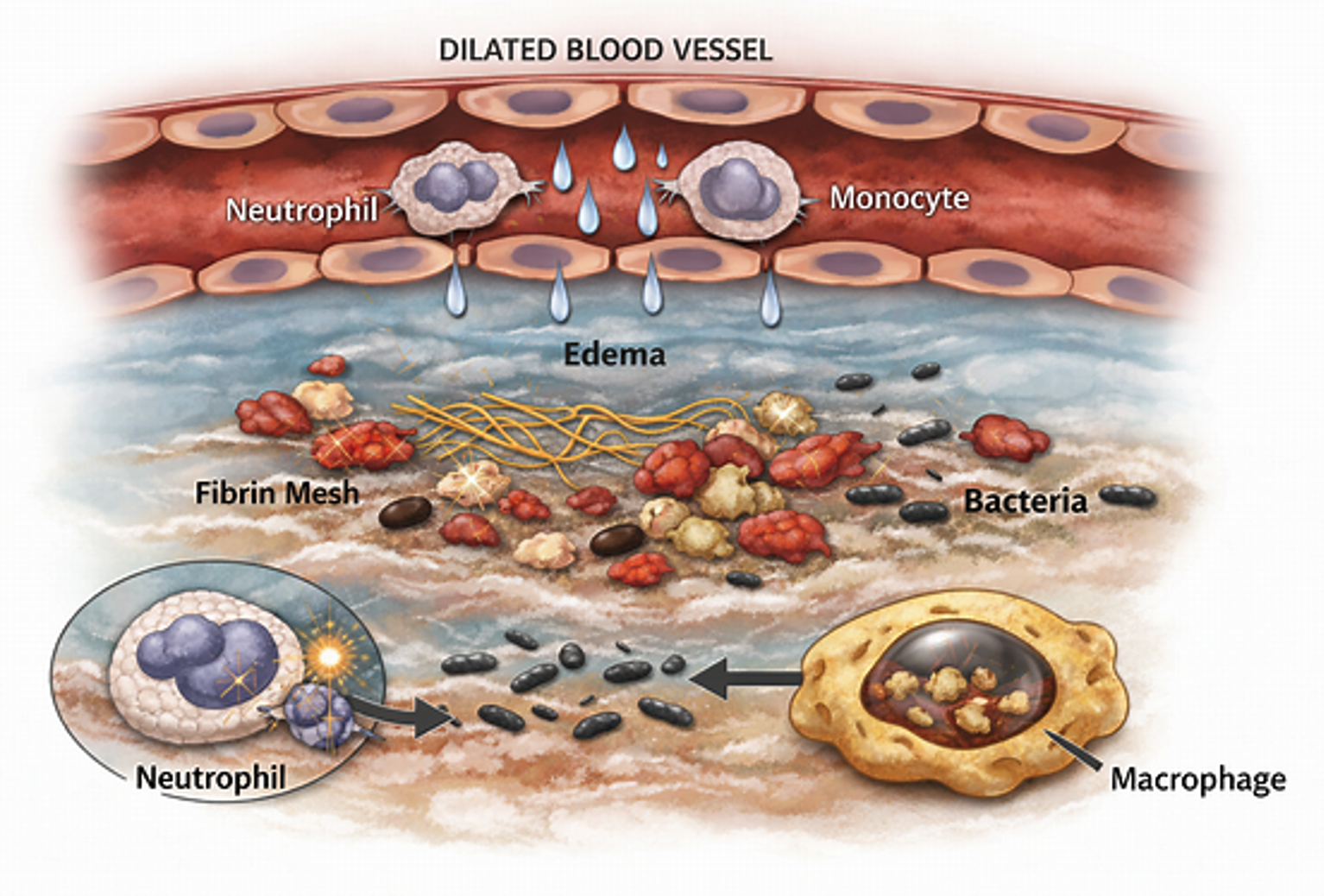
Intact endothelium normally releases nitric oxide (NO) and prostacyclin (PGI₂), which maintain smooth muscle relaxation and inhibit platelet activation. Within seconds to minutes after injury, damaged endothelial cells stop producing NO and PGI₂ and instead release endothelin‑1, a strong vasoconstrictor. Endothelin‑1 binds to endothelin A (ETA) receptors on vascular smooth muscle, causing strong contraction to limit blood loss until the platelet plug and fibrin clot form, and the loss of NO and PGI₂ removes inhibition on platelets, allowing collagen exposure to trigger their adhesion and aggregation. Exposure of collagen activates platelets through the surface receptors glycoprotein VI (GPVI) and integrin α2β1, which together stabilize platelet binding to collagen and anchor the growing platelet plug to the injured vessel wall. Activated platelets change shape—from discoid to spiky—via actin reorganization, and their granules release their contents; this includes serotonin, thromboxane A₂ (TXA₂), cytokines (e.g., IL‑1β, TNF‑α), CXCL and CCL chemokines, and growth factors (e.g., PDGF, TGF‑β, VEGF). Serotonin and TXA₂ contribute to local vasoconstriction, and sympathetic nerves in the vessel wall add to it further by releasing norepinephrine. TXA₂ also acts through a specific TP receptor on platelets which helps recruit nearby platelets by encouraging them to activate and move toward the growing platelet cluster. These combined actions allow platelets to rapidly accumulate at the injury site to form the initial soft platelet plug that slows bleeding before the fibrin clot develops. Platelet binding to collagen through GPVI and integrin α2β1 also activates integrin αIIbβ3 on the platelet surface. Activated integrin αIIbβ3 serves as the fibrinogen receptor, allowing plasma fibrinogen to attach to it and in so doing bridge adjacent platelets to each other to create true aggregation. Once the platelet plug is established, tissue factor exposed in the damaged vessel wall initiates the coagulation cascade, generating thrombin. Thrombin converts platelet‑bound fibrinogen into fibrin strands, and factor XIII crosslinks these strands to form a stable fibrin mesh. Plasma and cellular fibronectin co‑assemble with fibrin during polymerization, reinforcing the provisional matrix and creating additional integrin‑binding sites for platelets and leukocytes. As the fibrin–fibronectin scaffold matures, it interlaces the platelet plug and anchors the clot to the vessel wall, producing a firm, durable barrier that fully arrests bleeding. As hemostasis progresses to mature fibrin clot formation, the response remains confined to the site of vascular injury because nearby intact endothelium releases NO, PGI₂, and other inhibitors, while plasma anticoagulants such as antithrombin III and related factors limit further thrombin generation. This spatial confinement ensures that platelet activation and fibrin deposition do not propagate beyond the damaged surface. Late in hemostasis, transition to the inflammation stage takes place as endothelial permeability increases and leukocyte (neutrophil/monocyte) recruitment begins. Hemostasis contributes only limited initiating signals: platelets release a small set of vasoactive and matrix‑modifying factors, and thrombin provides early endothelial activation. These inputs prime the vascular interface but do not generate the main inflammatory drive. The tissue injury itself rapidly activates local inflammatory mechanisms—through resident sentinel cells (local injury‑sensing cells such as mast cells which release histamine), plasma cascades (injury‑activated plasma systems such as complement and bradykinin), and early leukocyte responses (chemokines, MMPs, serine proteases, and other short‑acting molecules)—which supply the dominant permeability and guidance cues for neutrophil and monocyte entry. As fibrin and the ECM undergo controlled proteolysis from plasmin and other plasma‑derived proteases, from platelet granule proteases, and from early leukocyte proteases, matrikines are generated that enhance leukocyte chemotaxis, while protease inhibitors maintain balance and prevent premature clot breakdown. The mature fibrin meshwork forms a three‑dimensional polymer that traps blood cells, microbes, and soluble mediators. This physical scaffold confines inflammatory signaling molecules—cytokines, chemokines, and growth factors—and retains recruited leukocytes at the wound surface, ensuring efficient progression into the inflammatory phase. Inflammation (3 hours to 2-5 days) As the fibrin clot mechanically limits blood loss, endothelial permeability beneath it increases because the endothelial cell–cell junctions physically loosen in response to thrombin, VEGF, and inflammatory cytokines (e.g., TNF‑α, IL‑1β, IL‑6). Endothelial cells at the same time increase the surface adhesion molecules required for neutrophil tethering, rolling, firm adhesion, and migration across the vessel wall, marking the transition of the clot‑covered wound into the inflammatory phase. In addition, platelets, damaged cells, and the remodeling provisional matrix release chemokines (e.g., CXCL1, CXCL2, CXCL8) and other short‑range inflammatory mediators (e.g., histamine, serotonin) that create local differences in concentration—stronger signals close to the wound and weaker signals farther away—which give neutrophils a clear sense of direction and guide them into the fibrin scaffold. With neutrophils now concentrated within the wound, they become activated by locally high levels of danger signals (short‑range molecular cues indicating tissue damage and acute inflammation) and begin clearing microbes and dead tissue through phagocytosis and enzyme release. These activated neutrophils release cytokines (e.g., TNF‑α, IL‑1β, IL‑6), chemokines (e.g., CXCL1/CXCL2, CCL2), and growth factors (e.g., TGF‑β, VEGF). Through their specific receptors, these molecular signals draw in additional leukocytes and activate nearby stromal cells—fibroblasts and other connective‑tissue support cells—as well as endothelial cells. These activated stromal and endothelial cells then release their own cytokines, chemokines, and growth factors, further intensifying the inflammatory response. This is how inflammation snowballs. As neutrophils complete their functions, degranulate, and undergo apoptosis, the signals they leave behind shift leukocyte entry from neutrophils to monocytes. This shift occurs because apoptotic neutrophils and activated stromal cells reduce their release of neutrophil‑directing CXCL chemokines and increase monocyte‑directing CCL chemokines—especially CCL2—which re‑targets leukocyte recruitment from neutrophils to monocytes. Monocytes enter the wound slightly later and differentiate into macrophages, which dominate the remainder of the inflammatory stage. Macrophages continue debris clearance but also perform essential regulatory functions. They release cytokines that shape the inflammatory environment—TNF‑α and IL‑1β to maintain controlled leukocyte recruitment, IL‑6 to support matrix remodeling, IL‑10 to limit excessive inflammation, and TGF‑β to begin shifting the wound from demolition toward reconstruction. Their growth factor release—particularly PDGF, TGF‑β, and VEGF—helps guide fibroblast migration, formation of new capillaries, and keratinocyte activation. Keratinocytes are the epithelial cells of the epidermis that later proliferate and migrate to restore the surface barrier. Through these actions, macrophages begin converting the fibrin‑rich clot into a biologically active scaffold that supports the transition into the proliferative phase. By the end of the inflammation stage, the wound has been cleared of debris, the provisional matrix has been remodeled into a structure that supports cell migration and new vessel growth, and the signaling environment has been reshaped to favor fibroblast, endothelial, and keratinocyte entry. These coordinated mechanisms ensure that proliferation begins only after the wound has been properly prepared, preserving the functional sequence required for successful healing (Figure 3).
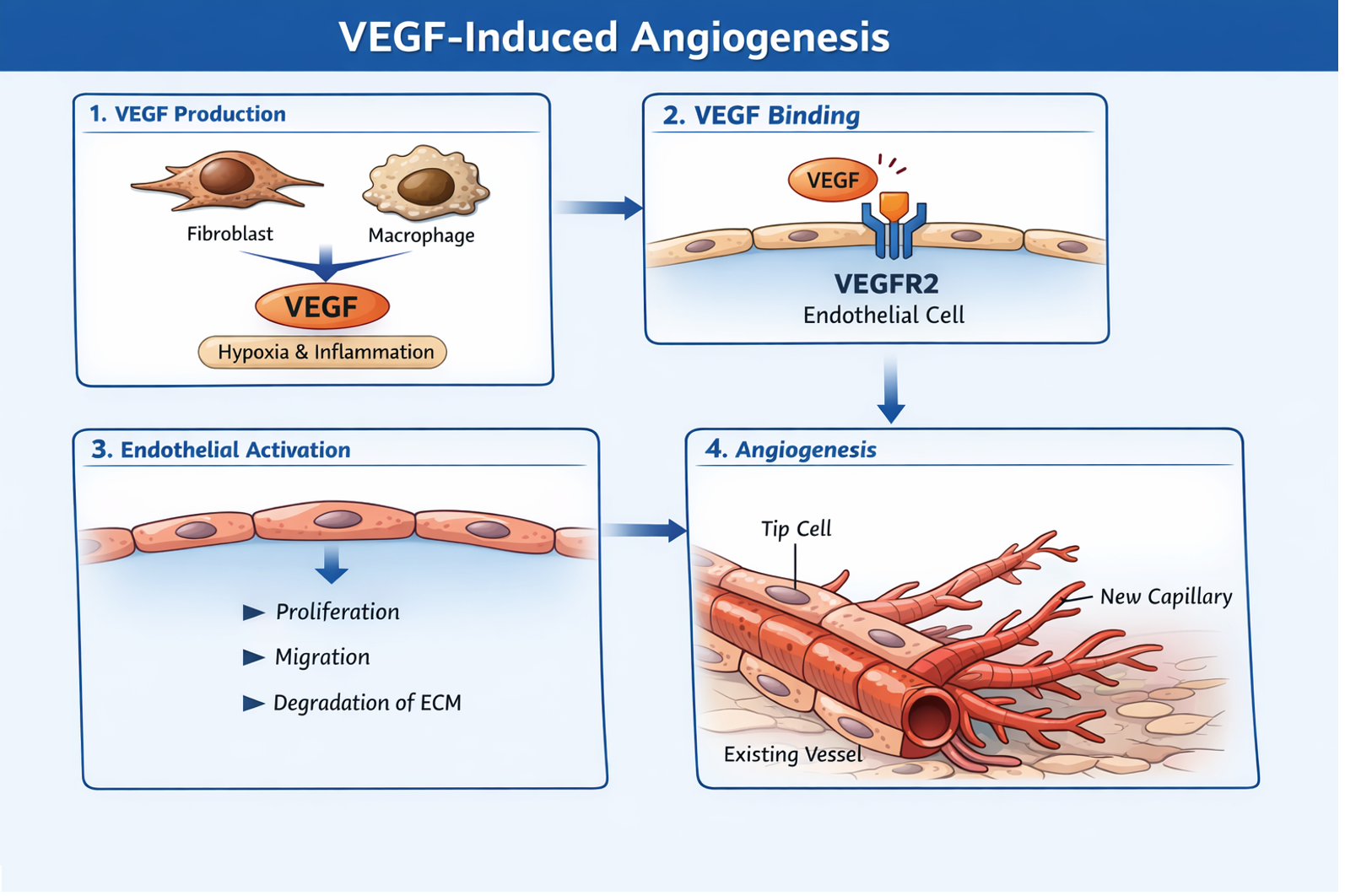
Proliferation (3 days to 21 days) Below the scab, early in the proliferative stage, fibroblasts from the wound margin migrate into the fibrin–fibronectin scaffold under the influence of growth factors (e.g., PDGF, TGF‑β) and chemokines (e.g., CCL2) released by platelets and macrophages. Through their specific receptors, these signals activate intracellular programs that ready the fibroblasts for movement—switching them into a motile state and enabling their entry into the wound. During migration, integrins on the surface of fibroblasts attach to fragments of collagens, fibronectin, and basement membranes. The low tension generated by these early adhesions produces mechanically derived signals that support forward movement. At the same time, matrix‑remodeling enzymes (e.g., MMPs, serine proteases) released from macrophages and fibroblasts clear away fibrin and damaged ECM, creating a pathway for fibroblasts to reach the fibrin–fibronectin scaffold. Once fibroblasts establish high‑tension attachments to the fibrin–fibronectin scaffold, the same growth factors and chemokines that initiated migration shift their effects on the fibroblasts to have them increase matrix production. Through changes in intracellular signaling, fibroblasts they begin to synthesize collagens, fibronectin, hyaluronan, and proteoglycans. This marks the beginning of the proliferative phase of matrix deposition. It is important to note here how the same signaling molecules can affect fibroblast behavior differently, depending on the context: whether the fibroblasts are just leaving the wound edge or have already attached to the fibrin–fibronectin scaffold. From here on, controlled ECM turnover is maintained by the balanced activity of MMPs and their inhibitors (TIMPs). This balance allows limited degradation of damaged ECM while preventing excessive breakdown, enabling fibroblasts to remodel the environment without destabilizing the developing provisional matrix. While fibroblasts are beginning matrix production, low oxygen (hypoxia) within the wound causes fibroblasts and macrophages to increase their release of VEGF. VEGF, together with other endothelial‑activating signals (e.g., PDGF, IL‑1β, TNF‑α), activates endothelial cells in nearby capillaries at the wound margin. Once activated, these endothelial cells loosen their junctions, reorganize their cytoskeleton, and initiate sprouting behavior by forming nascent endothelial extensions. At the same time, matrix‑remodeling enzymes (e.g., MMP‑2, MMP‑9) and plasmin open pathways through the provisional matrix, allowing these sprouting endothelial extensions to advance. The activated endothelial cells then proliferate and extend into the wound bed, preserving the integrity of the parent vessel while establishing the new microvasculature that will supply the proliferating tissue. Pericytes, which sit along the outer surface of capillaries, are subsequently recruited by PDGF to stabilize the newly formed vessels by supporting and strengthening their walls (Figure 4).
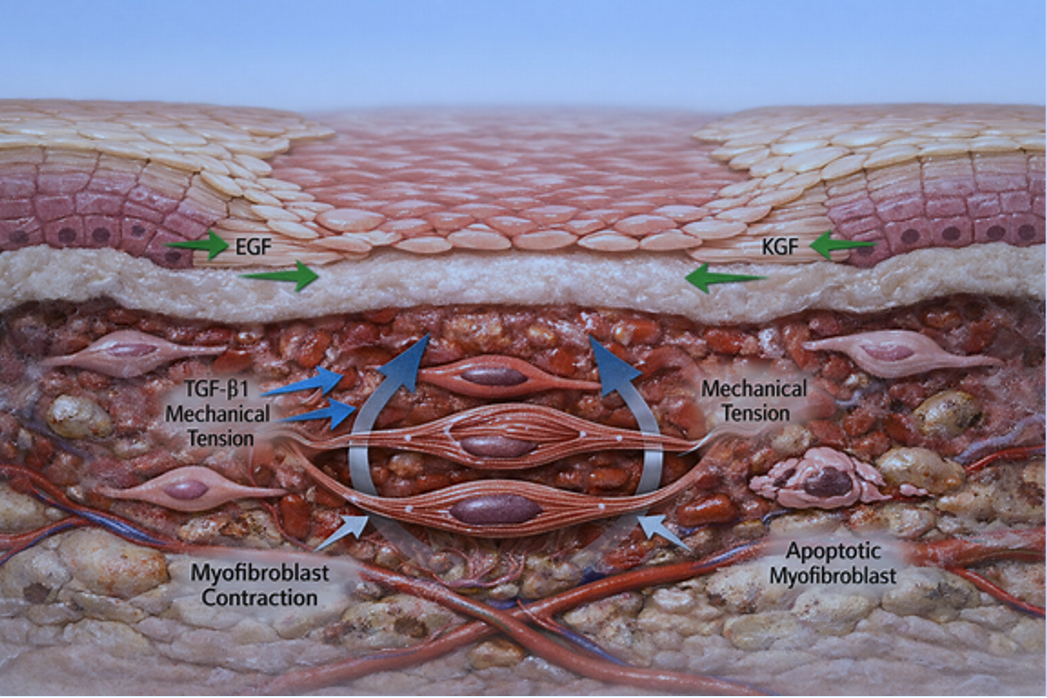
As new capillary sprouts enter the wound bed, fibroblasts continue migrating into the area and produce collagen, proteoglycans, and other ECM components. These elements collectively form the vascular, fibroblast‑rich provisional tissue known as granulation tissue. It fills the wound space and provides a temporary structural framework for cell migration. This process is supported by signaling from growth factors (e.g., PDGF, TGF-β), cytokines (e.g., IL-1β, At the wound edges, keratinocytes, the primary epidermal cells responsible for re‑establishing the skin barrier, loosen their attachments and begin migrating across the newly formed granulation tissue. Their movement is supported by Epidermal Growth Factor (EGF) and Keratinocyte Growth Factor (KGF) released from fibroblasts and macrophages. Keratinocytes move over the provisional matrix to re‑cover the wound surface. As they advance, they continue to divide, forming a new epithelial layer that gradually thickens. Once this new epithelial layer has sealed the wound surface, the fibrin clot loses its attachment to the underlying tissue and is shed. This restores the barrier function of the skin and protects the underlying tissue as healing continues. Within the granulation tissue, some fibroblasts differentiate into myofibroblasts, specialized contractile fibroblasts that generate tension within the wound. This transition is driven mainly by wound-specific TGF-β1 and mechanical forces within the matrix. Once formed, myofibroblasts use their contractile elements to pull the wound edges toward each other, reducing wound size and helping close the defect more efficiently. They remain active until sufficient contraction has occurred, after which many undergo apoptosis as the tissue progresses toward remodeling (Figure 5).
As the proliferative phase comes to an end, the intensity of cellular activity within the wound diminishes. Fibroblast proliferation slows, vascular density decreases, and the provisional matrix becomes progressively more organized. These shifts signal entry into the maturation/remodeling stage, during which collagen is reorganized, excess cells are removed, and the wound gradually gains tensile strength as the tissue moves toward its final structural form. Maturation/Remodeling (3 weeks to 12 months or more) As the wound enters the maturation/remodeling stage, the provisional matrix laid down during proliferation begins to be reorganized. The growth factors, cytokines, chemokines, enzymes, and enzyme inhibitors that were elevated earlier to drive inflammation, fibroblast recruitment, angiogenesis, and matrix deposition now decline overall as the wound shifts from active construction to controlled refinement. Fibroblasts reduce production of collagen, proteoglycans, and hyaluronan, and instead shift toward modifying and tightening the existing matrix, a change driven by a general downshift in their intracellular activity as the wound transitions out of the proliferative phase. This shift is influenced by continued low‑level signaling from TGF‑β1 and mechanical tension within the tissue, both of which help regulate fibroblast activity during this stage. Declining levels of PDGF and IL‑6 further reduce fibroblast proliferation, reinforcing the transition from matrix deposition to matrix refinement. Matrix turnover continues, but at a slower and more controlled rate. MMPs and TIMPs remain active, working together to remove damaged or excess matrix while preserving the newly formed collagen network. As collagen organization improves, overall MMP activity gradually decreases while TIMP activity maintains sufficient inhibitory tone to prevent excessive degradation. This balanced remodeling environment allows early type III collagen to be progressively replaced with stronger type I collagen. Increasing activity of collagen‑stabilizing enzymes strengthens intermolecular bonds within the matrix, and fibroblasts align collagen fibers along lines of mechanical stress, increasing tensile strength over time. As the matrix becomes more organized, fibroblasts gradually decrease in number. Many undergo apoptosis as their role diminishes, leaving behind a more stable collagen framework. Myofibroblasts also withdraw during this stage. Their contractile activity decreases as mechanical tension in the wound lessens, and most undergo apoptosis once contraction is complete. Reduced TGF‑β1 signaling contributes to this decline in myofibroblast activity, and the broader loss of pro‑survival cues—such as PDGF, IL‑6, and matrix tension—further promotes apoptosis in both fibroblasts and myofibroblasts. As these external signals fall and the matrix becomes mechanically stable, these cells lose the environmental support needed to persist, leading to their programmed removal. This reduction in cellularity is a key feature of the maturation/remodeling phase. The microvasculature formed during proliferation also undergoes refinement. As oxygen tension improves and inflammatory signals fall, fibroblasts become less responsive to VEGF. They express fewer VEGF‑binding receptors, rely less on matrix‑derived cues that previously heightened growth‑factor sensitivity, and no longer receive strong pro‑angiogenic support from macrophages and keratinocytes. At the same time, VEGF levels themselves decline as hypoxia resolves and the demand for new vessel formation decreases. Together, these changes limit fibroblast participation in angiogenesis and support the transition toward vascular stabilization and selective vessel regression. Pericytes remain attached to the vessels that persist, contributing to long‑term stability. The final result is a smaller, more efficient vascular network that matches the needs of the healed tissue. Keratinocytes continue to mature during this stage. The newly formed epithelial layer thickens and strengthens, restoring full barrier function. Growth factors such as EGF and KGF decline as the epithelial surface becomes stable, and keratinocyte proliferation slows to normal levels. Reduced IL‑1β and TNF‑α signaling further supports the transition from active epithelial repair to maintenance. Over the next several weeks to many months, collagen fibers become increasingly organized, cross‑linked, and aligned. The scar gradually becomes flatter, paler, and less cellular. Although the remodeled tissue never fully regains the strength of uninjured skin, nor its elasticity, it becomes significantly stronger and more stable than during earlier phases of healing. AI-Generated Evolutionary “Explanation” In Darwinian terms, human wound healing arose because undirected genetic variation produced small differences in the basic responses to injury—platelet adhesion and fibrin formation, neutrophil and macrophage activation through IL‑1, TNF‑α, and IL‑6, fibroblast activation through TGF‑β, keratinocyte proliferation for epithelial closure, collagen deposition, and VEGF‑driven angiogenesis. Primitive versions of these processes already existed and were low‑efficiency but viable. Individuals whose variants produced faster clotting, more effective inflammation, more reliable fibroblast activation, quicker epithelial repair, stronger collagen deposition, and more responsive angiogenesis survived injuries more often. Over many generations, these independently beneficial modules accumulated and stabilized through biochemical crosstalk and feedback. Modern human wound healing is simply the subset of parameter combinations that consistently supported survival; it appears coordinated because only coordinated variants persisted. Questions
|